|
||
| 18. Eye | ||
| 1 2 3 4 5 6 7 8 9 10 11 12 13 14 15 16 17 18 19 20 21 22 23 24 25 | ||
| 26 27 28 29 |
| |||
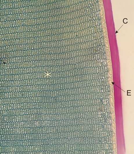 |
Section of the lens of a mouse stained with TPA and PAS.
This field presents the anterior surface of the lens. It shows the PAS positive capsule (C) equivalent to a thick basement membrane. Underlying the capsule, the anterior epithelium (E) of the lens is unstained and barely visible. The lens fibres (*) are cut transversely and their regular hexagonal profiles, well outlined in green by TPA, emphasize the regularity of their structure and packing. Stain: PAS-TPA
|
||