|
||
| 18. Eye | ||
| 1 2 3 4 5 6 7 8 9 10 11 12 13 14 15 16 17 18 19 20 21 22 23 24 25 | ||
| 26 27 28 29 |
| |||
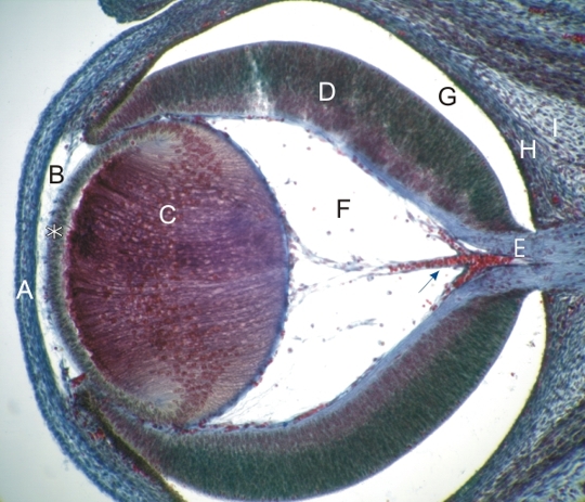 |
Section of the eye in a mouse embryo. This stage of eye development of a mouse corresponds to that of a human embryo of 6–7 weeks. The following structures can be recognized:
The area to be eventually occupied by the vitreous body (F) shows small vessels (arrow) branching from the vessel located in the centre of the optic nerve. The retina (D) is separated by the lumen of the embryonic optic vesicle (G) from the connective tissue that will form the choroids (H) and the sclera (I). The ciliary body and the iris are not yet formed at this stage of growth. Stain: Massons Trichrome
|
||