Contents Yves Clermont, Michael Lalli, Zsuzsanna Bencsath-Makkai
LIGHT-MICROSCOPIC HISTOLOGY ATLAS
     18. Eye
  1     2     3     4     5     6     7     8     9     10     11     12     13     14     15     16     17     18     19     20     21     22     23     24     25  
  26     27     28     29  

    18.14   Ciliary body Version française
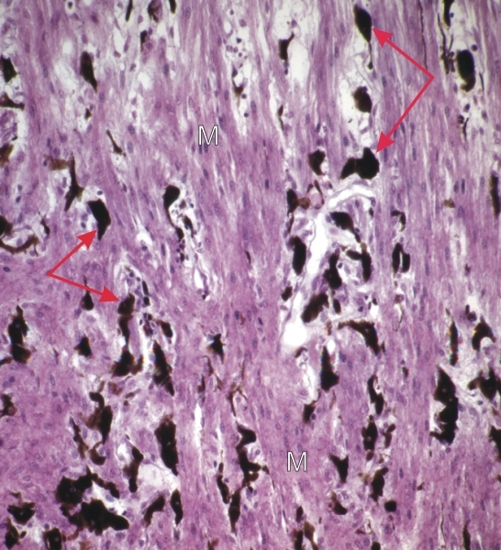
This section of the ciliary body shows bundles of smooth muscle fibres (M) separated by loose connective tissue containing numerous melanocytes (arrows).

These bundles of smooth muscle cells have two main orientations in the ciliary body. They are either longitudinal or circular, the circular ones being close to the ciliary processes (see Figure 18.10).

These ciliary muscles play an important role in visual accommodation.

Stain: H–­­E
Magnification: ×300


We'd very much appreciate receiving your comments and suggestions!We'd very much appreciate receiving your comments and suggestions!

Creative Commons Licence The text and images of this Histology Atlas, by Yves Clermont, Michael Lalli & Zsuzsanna Bencsath-Makkai, are licensed under a Creative Commons Attribution-Noncommercial-No Derivative Works 2.5 Canada Licence and cannot be modified without the written permission of the authors. Use of any text or images must carry an acknowledgement which includes a link to the original work.


HAweb/Zs B-M -> HAbase-generated on 2013-09-13