|
||
| 18. Eye | ||
| 1 2 3 4 5 6 7 8 9 10 11 12 13 14 15 16 17 18 19 20 21 22 23 24 25 | ||
| 26 27 28 29 |
| |||
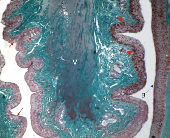 |
A dog's eyelid showing a fold of the conjunctiva from the bottom, or fornix of the eyelid.
This field corresponds to the framed area A in Figure 18.1. This fold shows the pseudostratified columnar epithelium in which some goblet cells secreting mucus are indicated (arrows). The epithelium on the right is partly pigmented (arrowheads). The epithelium covering these folds and the associated connective tissue is referred to as the palpebral conjunctiva. Underlying the palpebral conjunctiva, green-stained connective tissue (*) contains numerous small vessels (V). Facing this fold, the stratified squamous epithelium at the surface of the sclera and the underlying connective tissue belong to the bulbar conjunctiva (B). Stain: Massons Trichrome
|
||