Contents Yves Clermont, Michael Lalli, Zsuzsanna Bencsath-Makkai
LIGHT-MICROSCOPIC HISTOLOGY ATLAS
     18. Eye
  1     2     3     4     5     6     7     8     9     10     11     12     13     14     15     16     17     18     19     20     21     22     23     24     25  
  26     27     28     29  

    18.12   Ciliary body Version française
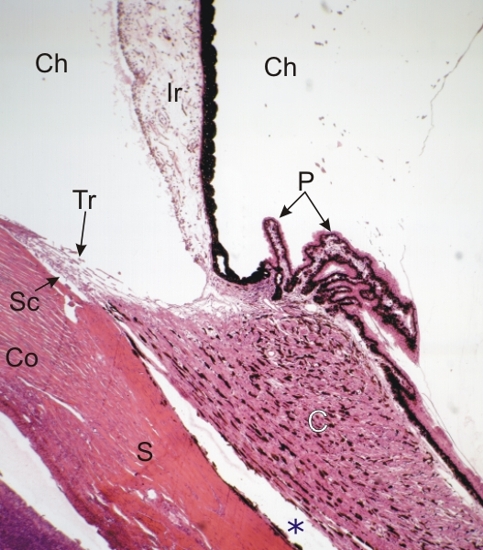
Section of the ciliary body of the eye. This field corresponds to the framed area B in Figure 18.1 and to Figure 18.10.

The following structures are labelled:

  • Sclera (S)
  • Ciliary body (C) composed of smooth muscles and melanocytes
  • Ciliairy processes (P) or fingerlike projections of the ciliary body
  • Iris (Ir) with a pigmented epithelium on its posterior surface.

Under the edge of the cornea (Co) the trabecular meshwork (Tr) and the canal of Schlemm (Sc) are visible. These two channels serve as exit routes for the aqueous humour present in the anterior and posterior chambers of the eye (Ch) Figure 18.13.

The space between the ciliary body and the sclera (*) is an artefact of tissue preparation.

Stain: H–­­E
Magnification: ×60


We'd very much appreciate receiving your comments and suggestions!We'd very much appreciate receiving your comments and suggestions!

Creative Commons Licence The text and images of this Histology Atlas, by Yves Clermont, Michael Lalli & Zsuzsanna Bencsath-Makkai, are licensed under a Creative Commons Attribution-Noncommercial-No Derivative Works 2.5 Canada Licence and cannot be modified without the written permission of the authors. Use of any text or images must carry an acknowledgement which includes a link to the original work.


HAweb/Zs B-M -> HAbase-generated on 2013-09-13