|
||
| 18. Eye | ||
| 1 2 3 4 5 6 7 8 9 10 11 12 13 14 15 16 17 18 19 20 21 22 23 24 25 | ||
| 26 27 28 29 |
| |||
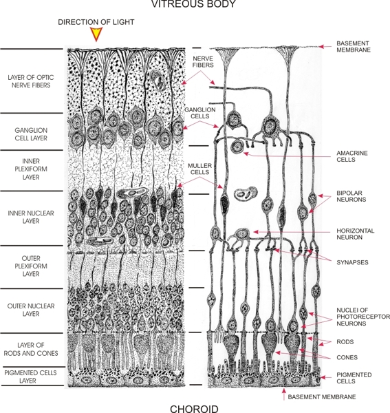 |
Drawing showing the components of the retina.
The various layers of the retina, as seen with the light microscope, are listed on the left, and the cells and their relations to each other are schematically represented on the right. The following neurones are illustrated:
The following neuroglia are also shown:
|
||