Contents Yves Clermont, Michael Lalli, Zsuzsanna Bencsath-Makkai
LIGHT-MICROSCOPIC HISTOLOGY ATLAS
     18. Eye
  1     2     3     4     5     6     7     8     9     10     11     12     13     14     15     16     17     18     19     20     21     22     23     24     25  
  26     27     28     29  

    18.16   Iris and lens Version française
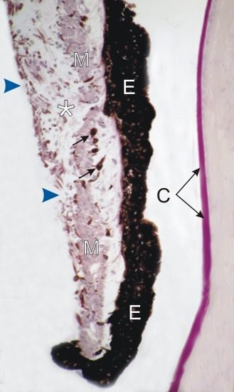
PAS-stained section of the iris and of the anterior surface of the lens on the right.

The following elements are identified:

  • Lightly stained cross sections of the smooth muscle cells (M) of the constrictor of the pupillae muscle
  • Loose connective tissue (*) containing some melanocytes (arrows)
  • Two layers of pigmented epithelial cells (E) on the posterior surface of the iris
  • Dicontinuous layer of connective tissue (arrowheads) on the anterior surface of the iris

(Right) The thick PAS-positive capsule of the lens (C) is equivalent to a thick basement membrane.

Stain: PAS-Hematoxylin
Magnification: ×225


We'd very much appreciate receiving your comments and suggestions!We'd very much appreciate receiving your comments and suggestions!

Creative Commons Licence The text and images of this Histology Atlas, by Yves Clermont, Michael Lalli & Zsuzsanna Bencsath-Makkai, are licensed under a Creative Commons Attribution-Noncommercial-No Derivative Works 2.5 Canada Licence and cannot be modified without the written permission of the authors. Use of any text or images must carry an acknowledgement which includes a link to the original work.


HAweb/Zs B-M -> HAbase-generated on 2013-09-13