|
||
| 18. Eye | ||
| 1 2 3 4 5 6 7 8 9 10 11 12 13 14 15 16 17 18 19 20 21 22 23 24 25 | ||
| 26 27 28 29 |
| |||
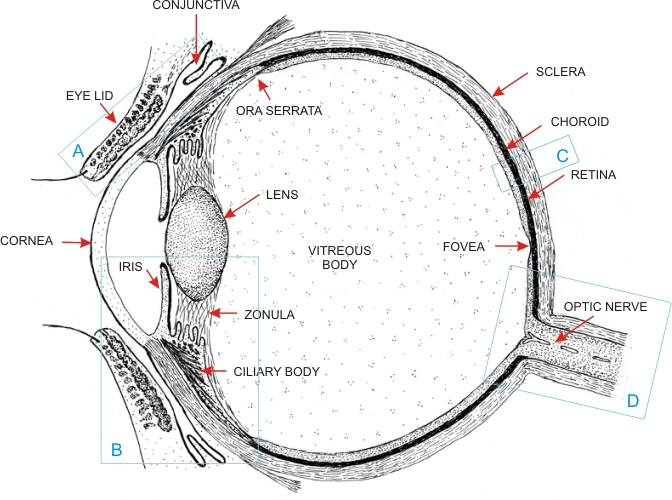 |
Drawing of the eye showing its main components, as seen in a sagittal section. The eyelids in front of the eyeball are also shown.
The cornea and the sclera of the eye, under the pressure of the aqueous humour and the vitreous body, maintain the shape of the eye. The cornea, the lens and the vitreous body constitute the transparent and refractive components of the eye. The iris, the ciliary body and the choroid form the protective and light-excluding components of the eye, owing to the presence of the melanin of pigmented cells. The retina, the photoreceptive layer of the eye, is connected to the optic nerve. The ciliary body and the associated zonular fibres are involved in the accommodation, or the focusing, of images on the retina. The framed areas A, B, C and D are examined in the following images:
|
||