Contents Yves Clermont, Michael Lalli, Zsuzsanna Bencsath-Makkai
LIGHT-MICROSCOPIC HISTOLOGY ATLAS
     18. Eye
  1     2     3     4     5     6     7     8     9     10     11     12     13     14     15     16     17     18     19     20     21     22     23     24     25  
  26     27     28     29  

    18.10   Eye Version française
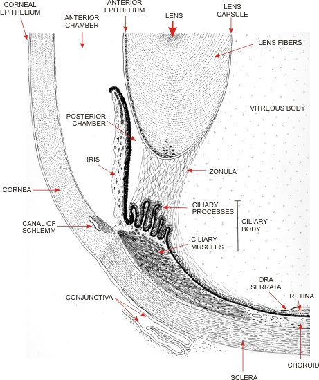
Drawing showing the anterior components of the ocular globe. This field corresponds to the framed area B in Figure 18.1.

The following structures are illustrated (they will be examined histologically in figures 18.11 to 18.24):

  • Cornea
  • Iris
  • Anterior and posterior chambers of the eye containing the aqueous humour
  • Ciliary body sitting on the sclera and composed of ciliary muscles and of fingerlike ciliary processes
  • Lens

The ciliary body is the insertion site of the zonular fibres that reach the lens and attach to its surface.

Posteriorly, the eye cavity is occupied by the transparent vitreous body.

The ciliary body is continuous posteriorly with the choroid and its epithelium is continuous with the retina at the ora serrata.


We'd very much appreciate receiving your comments and suggestions!We'd very much appreciate receiving your comments and suggestions!

Creative Commons Licence The text and images of this Histology Atlas, by Yves Clermont, Michael Lalli & Zsuzsanna Bencsath-Makkai, are licensed under a Creative Commons Attribution-Noncommercial-No Derivative Works 2.5 Canada Licence and cannot be modified without the written permission of the authors. Use of any text or images must carry an acknowledgement which includes a link to the original work.


HAweb/Zs B-M -> HAbase-generated on 2013-09-13