Contents Yves Clermont, Michael Lalli, Zsuzsanna Bencsath-Makkai
LIGHT-MICROSCOPIC HISTOLOGY ATLAS
     18. Eye
  1     2     3     4     5     6     7     8     9     10     11     12     13     14     15     16     17     18     19     20     21     22     23     24     25  
  26     27     28     29  

    18.3   Eyelids and cornea Version française
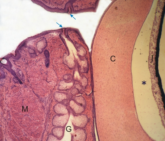
Anterior aspects of the eye of a monkey. (This section corresponds to the framed area A in Figure 18.1).

The edges of the eyelids are seen on the left and the cornea (C) and the iris (I) are seen on the right.

The external surface of the eyelid is lined with a stratified squamous epithelium. This field shows, at the margins of the lids, the openings of the ducts (arrows) of large sebaceous glands (G) called Meibomian glands.

In front of the large multilobular gland is a layer of striated muscle fibres (M).

The cornea is separated from the iris by the anterior chamber of the eye (*) containing the aqueous humour.

Stain: H–­­E
Magnification: ×10


We'd very much appreciate receiving your comments and suggestions!We'd very much appreciate receiving your comments and suggestions!

Creative Commons Licence The text and images of this Histology Atlas, by Yves Clermont, Michael Lalli & Zsuzsanna Bencsath-Makkai, are licensed under a Creative Commons Attribution-Noncommercial-No Derivative Works 2.5 Canada Licence and cannot be modified without the written permission of the authors. Use of any text or images must carry an acknowledgement which includes a link to the original work.


HAweb/Zs B-M -> HAbase-generated on 2013-09-13