Contents Yves Clermont, Michael Lalli, Zsuzsanna Bencsath-Makkai
LIGHT-MICROSCOPIC HISTOLOGY ATLAS
     18. Eye
  1     2     3     4     5     6     7     8     9     10     11     12     13     14     15     16     17     18     19     20     21     22     23     24     25  
  26     27     28     29  

    18.22   Lens Version française
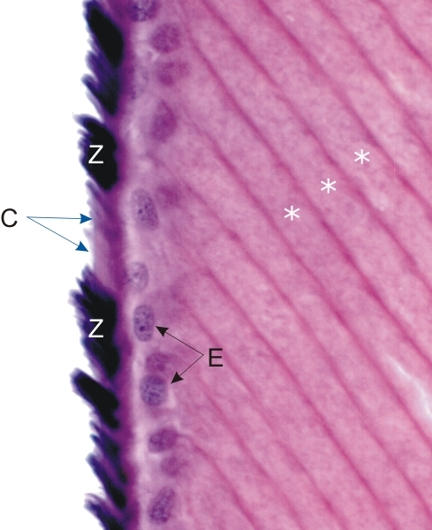
This field shows the anterior surface of the lens at its equator.

The lens fibres (*) terminate under the internal epithelium of the lens (E) underlying the lens capsule (C). These closely and regularly packed lens fibres are seen here cut longitudinally and running obliquely.

Numerous bundles of zonular fibres (Z), heavily stained black with iron hematoxylin, are deeply and obliquely inserted in the lens capsule (C).

Stain: Iron hematoxylin-E
Magnification: ×1000


We'd very much appreciate receiving your comments and suggestions!We'd very much appreciate receiving your comments and suggestions!

Creative Commons Licence The text and images of this Histology Atlas, by Yves Clermont, Michael Lalli & Zsuzsanna Bencsath-Makkai, are licensed under a Creative Commons Attribution-Noncommercial-No Derivative Works 2.5 Canada Licence and cannot be modified without the written permission of the authors. Use of any text or images must carry an acknowledgement which includes a link to the original work.


HAweb/Zs B-M -> HAbase-generated on 2013-09-13