Contents Yves Clermont, Michael Lalli, Zsuzsanna Bencsath-Makkai
LIGHT-MICROSCOPIC HISTOLOGY ATLAS
     18. Eye
  1     2     3     4     5     6     7     8     9     10     11     12     13     14     15     16     17     18     19     20     21     22     23     24     25  
  26     27     28     29  

    18.17   Iris Version française
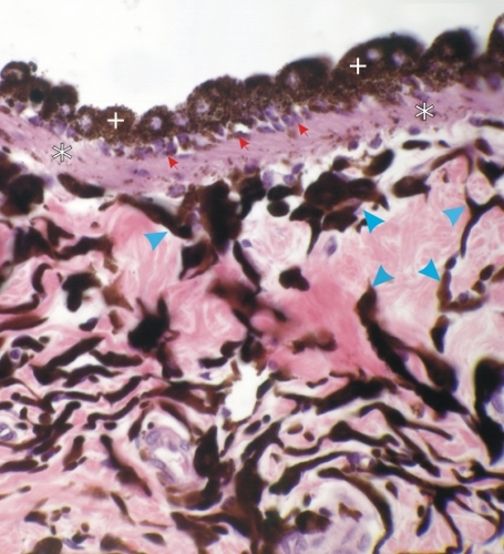
Section of a heavily pigmented iris of a dog.

In this field the iris contains numerous pigmented melanocytes (arrowheads) among the acidophilic collagen fibres.

The pigmented epithelium lining the posterior surface of the iris (top) shows two layers of pigmented cells: the large heavily pigmented cuboidal cells at the surface (+) and the deeper, less-pigmented myoepithelial cells (small red arrows).

These myoepithelial cells have long non-pigmented contractile cytoplasmic processes (*) arranged radially in the iris. Bundles of these contractile cellular processes form the dilator muscle of the pupil.

Stain: H–­­E
Magnification: ×300


We'd very much appreciate receiving your comments and suggestions!We'd very much appreciate receiving your comments and suggestions!

Creative Commons Licence The text and images of this Histology Atlas, by Yves Clermont, Michael Lalli & Zsuzsanna Bencsath-Makkai, are licensed under a Creative Commons Attribution-Noncommercial-No Derivative Works 2.5 Canada Licence and cannot be modified without the written permission of the authors. Use of any text or images must carry an acknowledgement which includes a link to the original work.


HAweb/Zs B-M -> HAbase-generated on 2013-09-13