|
||
| 18. il | ||
| 1 2 3 4 5 6 7 8 9 10 11 12 13 14 15 16 17 18 19 20 21 22 23 24 25 | ||
| 26 27 28 29 |
| |||
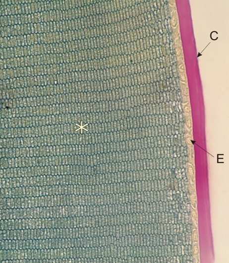 |
Coupe dun cristallin dune souris colorée au TPA et au PAS. Ce champ montre la surface antérieure du cristallin. La capsule (C), intensément colorée au PAS, correspond à une épaisse membrane basale. Lépithélium antérieur du cristallin (E) sous la capsule est incolore et à peine visible. Les fibres du cristallin sont ici coupées transversalement. Les limites hexagonales de ces fibres, bien délimitées par le TPA, soulignent la régularité de leur structure et de leur association dans le cristallin. Coloration: PAS-TPA
|
||