Sommaire Yves Clermont, Michael Lalli, Zsuzsanna Bencsath-Makkai
ATLAS D'HISTOLOGIE EN MICROSCOPIE OPTIQUE
     18. Œil
  1     2     3     4     5     6     7     8     9     10     11     12     13     14     15     16     17     18     19     20     21     22     23     24     25  
  26     27     28     29  

    18.22   Le cristallin English version
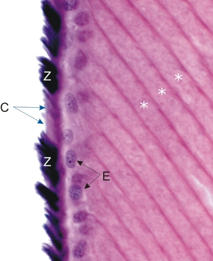
Ce champ montre la surface du cristallin à son equateur.

Cette photo montre des fibres du cristallin (*) coupées longitudinalement et disposées régulièrement côte à côte. Ces fibres du cristallin se terminent à la surface des cellules de l’épithélium antérieur du cristallin (E).

Des faisceaux des fibres de la zonule (Z), intensément colorées en noir par l’hématoxyline ferrique, sont profondément insérés obliquement dans la capsule du cristallin (C).

Coloration: Hématoxyline Ferrique-É
Grossissement: ×1000


We'd very much appreciate receiving your comments and suggestions!We'd very much appreciate receiving your comments and suggestions!

Creative Commons Licence The text and images of this Histology Atlas, by Yves Clermont, Michael Lalli & Zsuzsanna Bencsath-Makkai, are licensed under a Creative Commons Attribution-Noncommercial-No Derivative Works 2.5 Canada Licence and cannot be modified without the written permission of the authors. Use of any text or images must carry an acknowledgement which includes a link to the original work.


HAweb/Zs B-M -> HAbase-generated on 2013-09-13