|
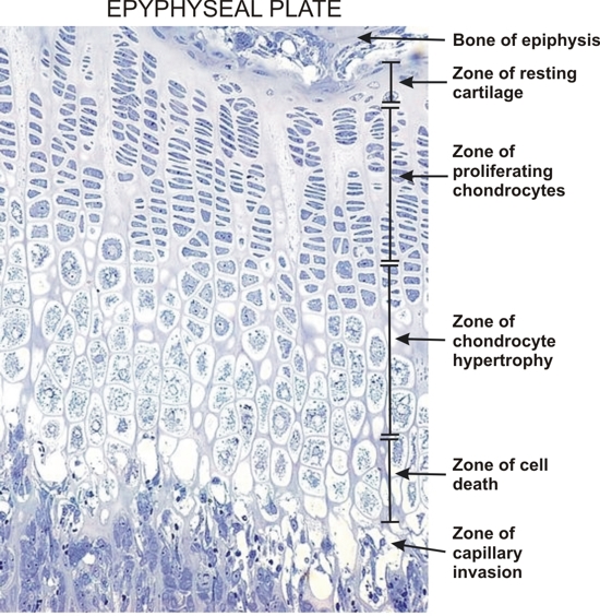
Section of the epiphyseal plate of a young rat.
Such a section was used to prepare the drawing in Figure 8.23. The various zones of the plate (top to bottom) illustrate the modifications of the chondrocytes during their evolution. Note that these modifications are gradual and therefore the boundaries of the zones are not sharp.
Following the degeneration of the chondrocytes in the zone of cell death, the spaces formed are occupied by capillaries and accompanying connective tissue cells.
Some of these differentiate into osteoblasts that deposit bone at the surface of the residual calcified cartilage matrix, thus producing mixed spicules.
Stain: Toluidine blue
Magnification: ×400
|