|
||
| 8. Cartilage and Bone | ||
| 1 2 3 4 5 6 7 8 9 10 11 12 13 14 15 16 17 18 19 20 21 22 23 24 25 | ||
| 26 27 28 29 30 31 |
| |||
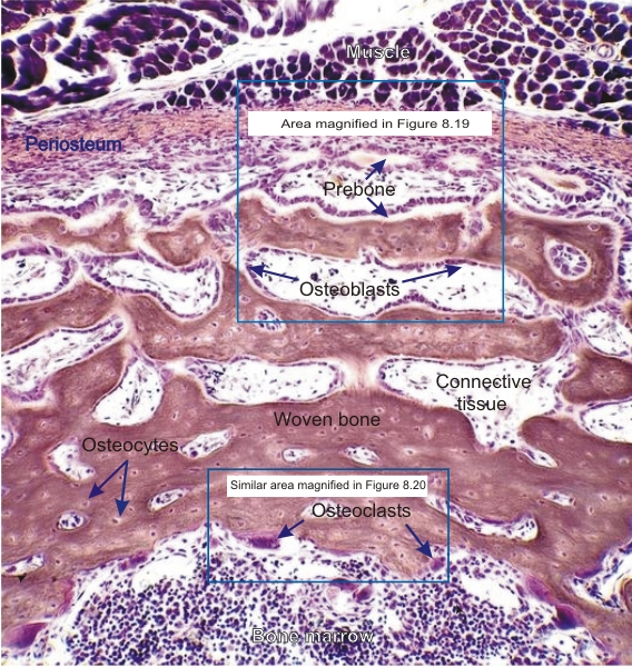 |
Diaphysis of a long bone of a cat embryo at the late foetal stage.
Compare with the other stages of growth illustrated in Figure 8.16. This field shows the periosteum underlying the skeletal muscle (top), and the bone marrow cavity (below). This growing diaphysis is composed of interconnected trabecules of primary (woven) bone separated by loose connective tissue. The trabecules on the periosteal side are widespread and show obvious signs of active osteoblastic bone formation. The osteoblasts on the endosteal side are rare and the primary bone spicules are more closely packed. In the bone marrow space, osteoclasts are seen along the bone. Through their lytic action, osteoblasts contribute to widening of the bone marrow cavity. Note that the framed areas are shown at a higher magnification in figures 8.19 and 8.20. Stain: Iron hematoxylin-E
|
||