|
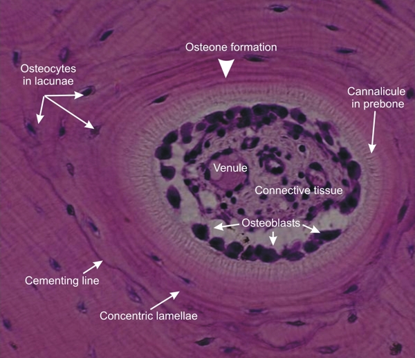
Osteon in reconstruction within the compact bone of an adult animal.
In the large central cavity of the osteon in formation, the loose connective tissue shows at its periphery a layer of intensely stained cells, the osteoblasts. These cells are forming the unmineralized pre-bone. In this pre-bone, the canaliculi are clearly visible.
This pre-bone calcifies to produce concentric lamellae and in the process some osteoblasts become embedded within the pre-bone and transform into osteocytes lodged within lacunae. This repetitive process leads to the formation of the concentric calcified bone lamellae of a new osteon.
Stain: HE
Magnification: ×1200
|