|
||
| 8. Cartilage and Bone | ||
| 1 2 3 4 5 6 7 8 9 10 11 12 13 14 15 16 17 18 19 20 21 22 23 24 25 | ||
| 26 27 28 29 30 31 |
| |||
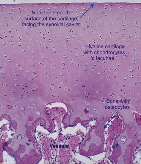 |
Hyaline cartilage at the articular extremity of a long bone of a young dog.
The synovial cavity is at the top and the epiphyseal marrow cavity with its vessels is at the base. The latter is lined with mixed trabecules. The hyaline cartilage shows clusters of chondrocytes. Vessels are absent from this hyaline cartilage. The articular surface, facing the synovial cavity, is smooth. Stain: HE
|
||