|
||
| 8. Cartilage and Bone | ||
| 1 2 3 4 5 6 7 8 9 10 11 12 13 14 15 16 17 18 19 20 21 22 23 24 25 | ||
| 26 27 28 29 30 31 |
| |||
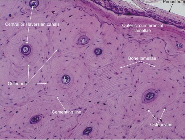 |
Compact bone from the periosteal side of a transversely cut diaphysis of a long bone from an adult dog. This is a higher magnification of the framed area in Figure 8.9.
At the top right of the field, the periosteum is seen at the surface of the outer circumferential lamellae. In the rest of the field, the bone is composed of lamellae, most of which are arranged concentrically around the central (Haversian) canals. These areas, called osteons, are composed of concentric lamellae forming cylinders oriented along the long axis of the long bones. The borderlines of the osteons are sometimes called cementing lines. The osteocytes lodged in small cavities or lacunae are arranged along the calcified bone lamellae. Stain: HE
|
||