Contents Yves Clermont, Michael Lalli, Zsuzsanna Bencsath-Makkai
LIGHT-MICROSCOPIC HISTOLOGY ATLAS
     8. Cartilage and Bone
  1     2     3     4     5     6     7     8     9     10     11     12     13     14     15     16     17     18     19     20     21     22     23     24     25  
  26     27     28     29     30     31  

    8.16   Stages of bone development Version française
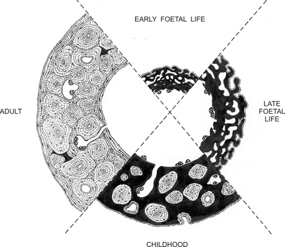
This drawing schematically illustrates the four stages of the radial growth of the diaphysis of a long bone. The primary bone is shown in black and the lamellar bone is pale and striated.

In early and late foetal life, bone is composed exclusively of trabecules of primary bone, also referred to as woven bone (black). The osteoblasts are shown as dots at the periphery of the superficial trabecules. Larger osteoclasts are shown on the endosteal surface. During childhood, concentric bone lamellae are formed in spaces present between trabecules of primary (woven) bone. This primary bone progressively disappears through the dissolving action of osteoclasts. In adults, compact bone is composed almost exclusively of lamellar bone, which is continually restructured by dissolution and replacement by osteon reconstruction as illustrated in figures 8.14 and 8.15.


We'd very much appreciate receiving your comments and suggestions!We'd very much appreciate receiving your comments and suggestions!

Creative Commons Licence The text and images of this Histology Atlas, by Yves Clermont, Michael Lalli & Zsuzsanna Bencsath-Makkai, are licensed under a Creative Commons Attribution-Noncommercial-No Derivative Works 2.5 Canada Licence and cannot be modified without the written permission of the authors. Use of any text or images must carry an acknowledgement which includes a link to the original work.


HAweb/Zs B-M -> HAbase-generated on 2013-09-13