Contents Yves Clermont, Michael Lalli, Zsuzsanna Bencsath-Makkai
LIGHT-MICROSCOPIC HISTOLOGY ATLAS
     8. Cartilage and Bone
  1     2     3     4     5     6     7     8     9     10     11     12     13     14     15     16     17     18     19     20     21     22     23     24     25  
  26     27     28     29     30     31  

    8.13   Compact bone Version française
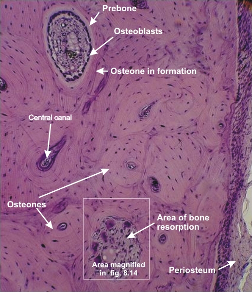
Compact bone of the diaphysis of a long bone of an adult dog.

This field shows the outer circumferential lamellae and the adjacent osteons with their respective central canals cut transversely or obliquely.

The framed area, shown at a higher magnification in Figure 8.14, illustrates an area of bone resorption.

Above is an osteon in reconstruction with osteoblasts at its periphery depositing the lightly stained pre-bone that will calcify to yield a concentric bone lamella.

Stain: H–­­E
Magnification: ×300


We'd very much appreciate receiving your comments and suggestions!We'd very much appreciate receiving your comments and suggestions!

Creative Commons Licence The text and images of this Histology Atlas, by Yves Clermont, Michael Lalli & Zsuzsanna Bencsath-Makkai, are licensed under a Creative Commons Attribution-Noncommercial-No Derivative Works 2.5 Canada Licence and cannot be modified without the written permission of the authors. Use of any text or images must carry an acknowledgement which includes a link to the original work.


HAweb/Zs B-M -> HAbase-generated on 2013-09-13