Contents Yves Clermont, Michael Lalli, Zsuzsanna Bencsath-Makkai
LIGHT-MICROSCOPIC HISTOLOGY ATLAS
     8. Cartilage and Bone
  1     2     3     4     5     6     7     8     9     10     11     12     13     14     15     16     17     18     19     20     21     22     23     24     25  
  26     27     28     29     30     31  

    8.30   Epiphyseal plate Version française
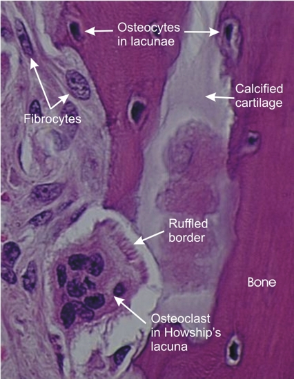
A mixed spicule associated with a large multinucleated osteoclast.

Owing to the fixation, this osteoclast is slightly retracted from the spicule. A part of the osteoclast faces a depression of the spicule called Howship’s lacuna. Facing the bone and the calcified cartilage of the spicule, the osteoclast shows a ruffled border or a row of cytoplasmic processes which release lytic substances that dissolve both recently deposited bone and calcified cartilage.

Stain: H–­­E
Magnification: ×1000


We'd very much appreciate receiving your comments and suggestions!We'd very much appreciate receiving your comments and suggestions!

Creative Commons Licence The text and images of this Histology Atlas, by Yves Clermont, Michael Lalli & Zsuzsanna Bencsath-Makkai, are licensed under a Creative Commons Attribution-Noncommercial-No Derivative Works 2.5 Canada Licence and cannot be modified without the written permission of the authors. Use of any text or images must carry an acknowledgement which includes a link to the original work.


HAweb/Zs B-M -> HAbase-generated on 2013-09-13