|
||
| 8. Cartilage and Bone | ||
| 1 2 3 4 5 6 7 8 9 10 11 12 13 14 15 16 17 18 19 20 21 22 23 24 25 | ||
| 26 27 28 29 30 31 |
| |||
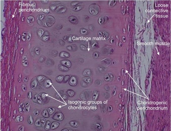 |
Cartilage plate of a small bronchus of a rodent.
This field shows numerous chondrocytes lodged in spheroidal or hemispherical cavities called lacunae. Chondrocytes are frequently arranged in clusters called isogenic groups, which are formed during growth. On the two surfaces of the cartilage plate, the osteoblasts are small and flattened, and form a layer called the chondrogenic perichondrium. External to this layer, the loose connective tissue is composed of collagen and fibrocytes called fibrous perichondrium. Stain: HE
|
||