Contents Yves Clermont, Michael Lalli, Zsuzsanna Bencsath-Makkai
LIGHT-MICROSCOPIC HISTOLOGY ATLAS
     8. Cartilage and Bone
  1     2     3     4     5     6     7     8     9     10     11     12     13     14     15     16     17     18     19     20     21     22     23     24     25  
  26     27     28     29     30     31  

    8.28   Epiphyseal plate Version française
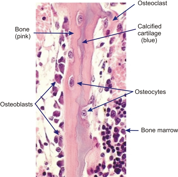
A mixed spicule from the underlying aspect of an epiphyseal plate.

On one side of the spicule is a layer of osteoblasts lining the newly formed bone. Osteocytes, which derive from osteoblasts, are seen at various stages of incorporation into their respective lacunae.

A small amount of calcified cartilage is visible in the core of the spicule.

Cells of the bone marrow are indicated.

Stain: H–­­E
Magnification: ×600


We'd very much appreciate receiving your comments and suggestions!We'd very much appreciate receiving your comments and suggestions!

Creative Commons Licence The text and images of this Histology Atlas, by Yves Clermont, Michael Lalli & Zsuzsanna Bencsath-Makkai, are licensed under a Creative Commons Attribution-Noncommercial-No Derivative Works 2.5 Canada Licence and cannot be modified without the written permission of the authors. Use of any text or images must carry an acknowledgement which includes a link to the original work.


HAweb/Zs B-M -> HAbase-generated on 2013-09-13