Contents Yves Clermont, Michael Lalli, Zsuzsanna Bencsath-Makkai
LIGHT-MICROSCOPIC HISTOLOGY ATLAS
     8. Cartilage and Bone
  1     2     3     4     5     6     7     8     9     10     11     12     13     14     15     16     17     18     19     20     21     22     23     24     25  
  26     27     28     29     30     31  

    8.4   Hyaline cartilage and perichondrium Version française
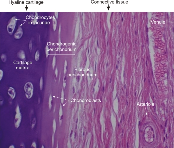
Higher magnification of the framed area in Figure 8.3 showing the edge of a cartilage plate.

In the hyaline cartilage, the poorly fixed chondrocytes in their lacunae are separated by the strongly basophilic matrix.

At the edge of this plate, the chondrogenic perichondrium shows the flattened chondroblasts embedded in a paler matrix.

The fibrous perichondrium is composed of fibrocytes and collagen fibres. The layer of collagen fibres is not sharply demarcated from the dense connective tissue composing the rest of the tracheal wall.

Stain: H–­­E
Magnification: ×400


We'd very much appreciate receiving your comments and suggestions!We'd very much appreciate receiving your comments and suggestions!

Creative Commons Licence The text and images of this Histology Atlas, by Yves Clermont, Michael Lalli & Zsuzsanna Bencsath-Makkai, are licensed under a Creative Commons Attribution-Noncommercial-No Derivative Works 2.5 Canada Licence and cannot be modified without the written permission of the authors. Use of any text or images must carry an acknowledgement which includes a link to the original work.


HAweb/Zs B-M -> HAbase-generated on 2013-09-13