Contents Yves Clermont, Michael Lalli, Zsuzsanna Bencsath-Makkai
LIGHT-MICROSCOPIC HISTOLOGY ATLAS
     8. Cartilage and Bone
  1     2     3     4     5     6     7     8     9     10     11     12     13     14     15     16     17     18     19     20     21     22     23     24     25  
  26     27     28     29     30     31  

    8.1   Growing long bone Version française
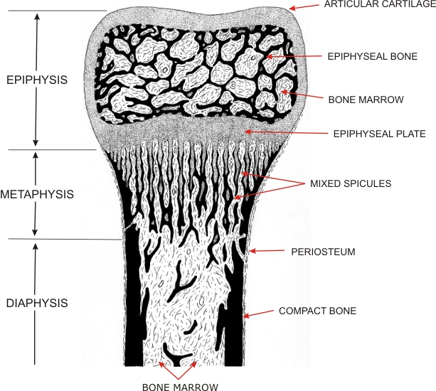
Drawing of a section of growing long bone to show the distribution of the typical calcified bone tissue (solid black) and of cartilage (grey).

In this growing bone, the calcified tissue forms the compact wall of the diaphysis around the bone marrow cavity as well as part of the spicules of the metaphysis.

The hyaline cartilage forms the core of the spicules of the metaphysis and the epiphyseal plate as well as the periphery of the epiphysis.


We'd very much appreciate receiving your comments and suggestions!We'd very much appreciate receiving your comments and suggestions!

Creative Commons Licence The text and images of this Histology Atlas, by Yves Clermont, Michael Lalli & Zsuzsanna Bencsath-Makkai, are licensed under a Creative Commons Attribution-Noncommercial-No Derivative Works 2.5 Canada Licence and cannot be modified without the written permission of the authors. Use of any text or images must carry an acknowledgement which includes a link to the original work.


HAweb/Zs B-M -> HAbase-generated on 2013-09-13