|
||
| 8. Cartilage and Bone | ||
| 1 2 3 4 5 6 7 8 9 10 11 12 13 14 15 16 17 18 19 20 21 22 23 24 25 | ||
| 26 27 28 29 30 31 |
| |||
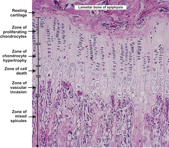 |
Epiphyseal plate of a long bone of a young dog. This field shows the epiphysis (top) and the metaphysis (bottom).
In addition to the radial growth of the diaphysis of a long bone, the growth in length of the bone takes place mainly at the level of the epiphyseal plate that separates the epiphyseal and diaphyseal cavities (see Figure 8.1). Underlying the lamellar bone of the epiphysis (top) is a thin layer of resting hyaline cartilage. Below, the various stages of chondrocyte transformation and of mixed spicule formation are labelled. The zones of the epiphyseal plate and the mode of mixed spicule formation are also schematically represented in Figure 8.23. Stain: HE
|
||