Contents Yves Clermont, Michael Lalli, Zsuzsanna Bencsath-Makkai
LIGHT-MICROSCOPIC HISTOLOGY ATLAS
     8. Cartilage and Bone
  1     2     3     4     5     6     7     8     9     10     11     12     13     14     15     16     17     18     19     20     21     22     23     24     25  
  26     27     28     29     30     31  

    8.7   Fibrocartilage Version française
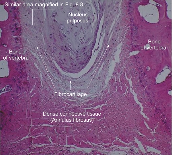
Section of an intervertebral disc from the tail of a dog.

On the extreme left and right of the image the edges of the two facing vertebrae are visible.

The upper central portion of the field is occupied by the nucleus pulposus, a gelatinous discoid mass surrounded by a layer of fibrocartilage (see the framed area in Figure 8.8).

More peripheral and below the layer of fibrocartilage the dense connective tissue of the annulus fibrosus, bridges the two vertebrae.

Stain: H–­­E
Magnification: ×50


We'd very much appreciate receiving your comments and suggestions!We'd very much appreciate receiving your comments and suggestions!

Creative Commons Licence The text and images of this Histology Atlas, by Yves Clermont, Michael Lalli & Zsuzsanna Bencsath-Makkai, are licensed under a Creative Commons Attribution-Noncommercial-No Derivative Works 2.5 Canada Licence and cannot be modified without the written permission of the authors. Use of any text or images must carry an acknowledgement which includes a link to the original work.


HAweb/Zs B-M -> HAbase-generated on 2013-09-13