 |
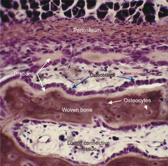
Diaphysis of a long bone of a cat on its periosteal side (see Figure 8.18).
This field shows the periosteum underlying the muscle (above) and the early stages of formation of bone spicules (below), with osteoblasts depositing lightly stained uncalcified pre-bone. This pre-bone later calcifies to become the more heavily stained primary (woven) bone, forming the spicules. Osteocytes, which derive from osteoblasts, are seen enclosed in the lacunae of this primary bone.
Stain: Iron hematoxylin-E
Magnification: ×300
|