Contents Yves Clermont, Michael Lalli, Zsuzsanna Bencsath-Makkai
LIGHT-MICROSCOPIC HISTOLOGY ATLAS
     10. Respiratory System
  1     2     3     4     5     6     7     8     9     10     11     12     13     14     15     16     17     18     19     20     21     22     23     24     25  
  26     27  

    10.8   Bronchus Version française
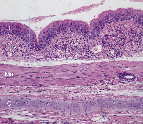
Section of the wall of a medium-sized bronchus of a dog.

This field shows the following components of the wall of bronchioles (top to bottom):

  • Pseudostratified columnar epithelium (E)
  • Lamina propria (LP)
  • A continuous layer of smooth muscle fibers (Mu)
  • A plate of hyaline cartilage (C) with layers of dense perichondrium on both surfaces of the plate
The framed area is shown at a higher magnification in Figure 10.9.

Stain: H–­­E
Magnification: ×250


We'd very much appreciate receiving your comments and suggestions!We'd very much appreciate receiving your comments and suggestions!

Creative Commons Licence The text and images of this Histology Atlas, by Yves Clermont, Michael Lalli & Zsuzsanna Bencsath-Makkai, are licensed under a Creative Commons Attribution-Noncommercial-No Derivative Works 2.5 Canada Licence and cannot be modified without the written permission of the authors. Use of any text or images must carry an acknowledgement which includes a link to the original work.


HAweb/Zs B-M -> HAbase-generated on 2013-09-13