Contents Yves Clermont, Michael Lalli, Zsuzsanna Bencsath-Makkai
LIGHT-MICROSCOPIC HISTOLOGY ATLAS
     10. Respiratory System
  1     2     3     4     5     6     7     8     9     10     11     12     13     14     15     16     17     18     19     20     21     22     23     24     25  
  26     27  

    10.11   Bronchus Version française
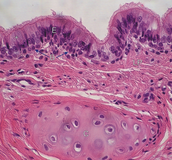
Section of a small bronchus.

A small piece of hyaline cartilage (C) characterizes this small bronchus. (N.B.: There is no cartilage in the wall of bronchioles. See Figure 10.12.)

The pseudostratified epithelium (E) shows ciliated cells, some goblet cells (+) and basal cells (B).

The lamina propria (LP) is also labelled.

Stain: H–­­E
Magnification: ×550


We'd very much appreciate receiving your comments and suggestions!We'd very much appreciate receiving your comments and suggestions!

Creative Commons Licence The text and images of this Histology Atlas, by Yves Clermont, Michael Lalli & Zsuzsanna Bencsath-Makkai, are licensed under a Creative Commons Attribution-Noncommercial-No Derivative Works 2.5 Canada Licence and cannot be modified without the written permission of the authors. Use of any text or images must carry an acknowledgement which includes a link to the original work.


HAweb/Zs B-M -> HAbase-generated on 2013-09-13