|
||
| 10. Respiratory System | ||
| 1 2 3 4 5 6 7 8 9 10 11 12 13 14 15 16 17 18 19 20 21 22 23 24 25 | ||
| 26 27 |
| |||
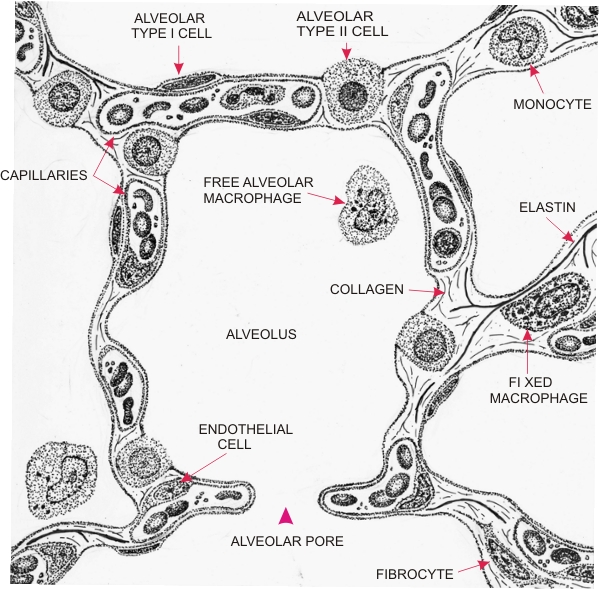 |
Drawing illustrating the composition of the wall of a pulmonary alveolus.
At the surface of the alveolar wall there are two types of epithelial cells: the squamous alveolar type I and the cuboidal alveolar type II cells. The latter cells secrete surfactant, a lipid substance that spreads along the surface of the epithelium and prevents the alveoli from collapsing. The capillaries constitute the major components of the alveolar wall. The following elements are also components of the alveolar wall: fibrocytes, monocytes, fixed macrophages, and reticular and elastic fibres. Alveolar type I and endothelial cells of capillaries are closely apposed to each other and at these sites they have a common basement membrane visible only with the electron microscope and not shown on this drawing. At such points the thin cytoplasmic layers of endothelial and of type I alveolar cells, plus the basement membrane, may be as small as 0.2 µ thick, which is at the limit of visibility of the light microscope. Such a fine partition permits the exchange of oxygen and carbon dioxide between air and red blood cells. Free-wandering alveolar macrophages of bone marrow origin are also present in the lumen of the alveoli. In addition to the broad openings between the alveoli (not shown here), there are smaller gaps or alveolar pores which serve as additional air passages between adjacent alveoli.
|
||