|
||
| 10. Respiratory System | ||
| 1 2 3 4 5 6 7 8 9 10 11 12 13 14 15 16 17 18 19 20 21 22 23 24 25 | ||
| 26 27 |
| |||
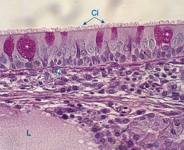 |
Section of tracheal mucosa.
The epithelium shows ciliated columnar cells (Ci) characterized by their lightly stained large nuclei. The mucous or goblet cells (+) are stained with PAS, owing to the carbohydrate-rich mucigen granules contained in their apical cytoplasm. The intensely stained small nuclei of basal epithelial cells are aligned along the basement membrane. The basement membrane (white arrow) is stained with PAS, owing to the carbohydrate-containing type IV collagen. The lamina propria contains a tracheal gland (G) and a lymphatic vessel (L), in addition to connective tissue cells and fibres. Stain: PAS-Hematoxylin
|
||