|
||
| 10. Respiratory System | ||
| 1 2 3 4 5 6 7 8 9 10 11 12 13 14 15 16 17 18 19 20 21 22 23 24 25 | ||
| 26 27 |
| |||
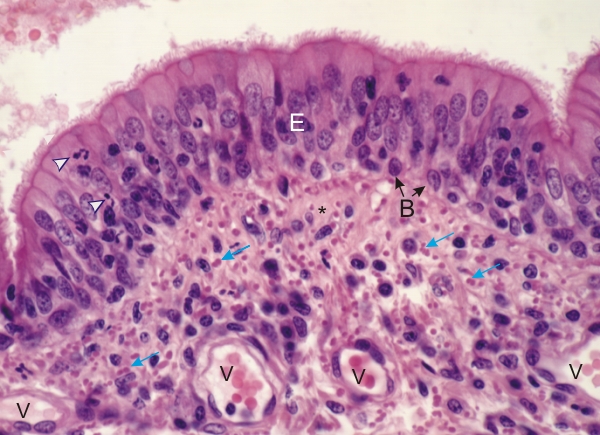 |
Section of the mucosa of a bronchus. This is a higher magnification of the framed area in Figure 10.8.
At the surface of the bronchus, the pseudostratified columnar epithelium (E) is lined with ciliated cells. It is invaded by leukocytes (arrowheads). Goblet cells are rare and difficult to identify. Basal cells (B) are labelled. The lamina propria contains many cells (fibrocytes and lymphoid cells) separated by pale pink collagen fibres (*) and many acidophilic elastic fibres seen here in cross section (arrows). Small blood vessels (V) are also labelled. Stain: HE
|
||