Contents Yves Clermont, Michael Lalli, Zsuzsanna Bencsath-Makkai
LIGHT-MICROSCOPIC HISTOLOGY ATLAS
     10. Respiratory System
  1     2     3     4     5     6     7     8     9     10     11     12     13     14     15     16     17     18     19     20     21     22     23     24     25  
  26     27  

    10.4   Trachea Version française
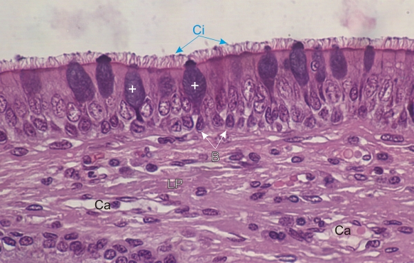
Section of the trachea of a dog.

This field shows the pseudostratified ciliated epithelium. It is composed of ciliated cells (Ci) with their large pale ovoid nuclei, of mucous goblet cells (+) with their basophilic mucigen granules and of basal cells (B).

In most preparations the highly soluble mucigen granules of goblet cells are extracted during the fixation of the tissue, leaving a lightly stained apical cytoplasm. In the present preparation the mucigen granules have been preserved and are intensely stained with hematoxylin.

The lamina propria (LP) composed of dense connective tissue shows some capillaries (Ca).

Stain: H–­­E
Magnification: ×500


We'd very much appreciate receiving your comments and suggestions!We'd very much appreciate receiving your comments and suggestions!

Creative Commons Licence The text and images of this Histology Atlas, by Yves Clermont, Michael Lalli & Zsuzsanna Bencsath-Makkai, are licensed under a Creative Commons Attribution-Noncommercial-No Derivative Works 2.5 Canada Licence and cannot be modified without the written permission of the authors. Use of any text or images must carry an acknowledgement which includes a link to the original work.


HAweb/Zs B-M -> HAbase-generated on 2013-09-13