Contents Yves Clermont, Michael Lalli, Zsuzsanna Bencsath-Makkai
LIGHT-MICROSCOPIC HISTOLOGY ATLAS
     10. Respiratory System
  1     2     3     4     5     6     7     8     9     10     11     12     13     14     15     16     17     18     19     20     21     22     23     24     25  
  26     27  

    10.17   Bronchiole Version française
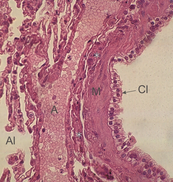
Section of the lung of a dog.

This field is a cross section of a respiratory bronchiole.

It shows an epithelium composed of Clara cells (Cl) sitting on a layer of smooth muscle fibres (M) cut longitudinally.

Underlying this muscular tunica is a layer of dense connective tissue (*) and beyond a longitudinal section of an arteriole (A).

Some collapsed alveoli (Al) are also visible on the left.

Stain: H–­­E
Magnification: ×800


We'd very much appreciate receiving your comments and suggestions!We'd very much appreciate receiving your comments and suggestions!

Creative Commons Licence The text and images of this Histology Atlas, by Yves Clermont, Michael Lalli & Zsuzsanna Bencsath-Makkai, are licensed under a Creative Commons Attribution-Noncommercial-No Derivative Works 2.5 Canada Licence and cannot be modified without the written permission of the authors. Use of any text or images must carry an acknowledgement which includes a link to the original work.


HAweb/Zs B-M -> HAbase-generated on 2013-09-13