|
||
| 10. Respiratory System | ||
| 1 2 3 4 5 6 7 8 9 10 11 12 13 14 15 16 17 18 19 20 21 22 23 24 25 | ||
| 26 27 |
| |||
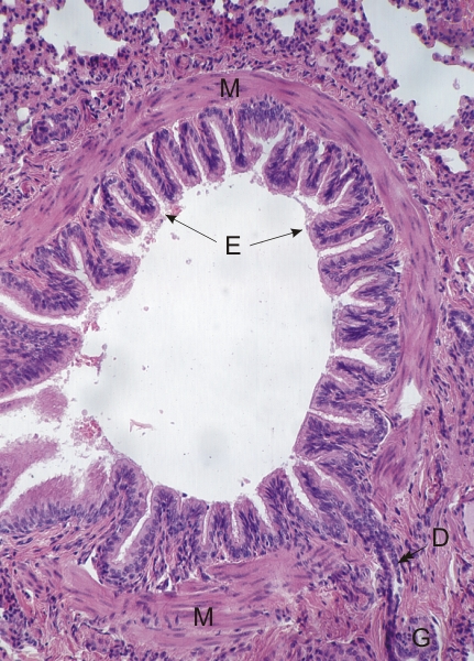 |
Section of a large bronchiole of a dog.
This air passage is lined with a pseudostratified ciliated columnar epithelium (E) covering folds of the mucosa. These folds are amplified here by the contraction of the tissues owing to the fixation of the lung. In the lamina propria and at a short distance from these folds there is a tunica composed of smooth muscle fibres (M). This muscular layer is crossed by the duct (D) of a small bronchiolar gland (G). Note that there is no cartilage in the walls of bronchioles. Stain: HE
|
||