Contents Yves Clermont, Michael Lalli, Zsuzsanna Bencsath-Makkai
LIGHT-MICROSCOPIC HISTOLOGY ATLAS
     10. Respiratory System
  1     2     3     4     5     6     7     8     9     10     11     12     13     14     15     16     17     18     19     20     21     22     23     24     25  
  26     27  

    10.16   Bronchiole Version française
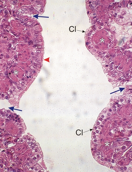
Section of the lung of a dog.

This field shows a respiratory bronchiole with a few connections (arrows) to the underlying alveoli. The respiratory bronchiole is lined with a simple epithelium composed of lightly stained cells, the Clara cells (Cl), showing a spherical nucleus and a slightly granulated cytoplasm. These cells secrete a serous fluid.

These Clara cells are next to bundles of smooth muscle fibres (*) seen here in cross section.

A few ciliated cells (arrowhead) are at the borderline of a terminal and a respiratory bronchiole.

Stain: H–­­E
Magnification: ×500


We'd very much appreciate receiving your comments and suggestions!We'd very much appreciate receiving your comments and suggestions!

Creative Commons Licence The text and images of this Histology Atlas, by Yves Clermont, Michael Lalli & Zsuzsanna Bencsath-Makkai, are licensed under a Creative Commons Attribution-Noncommercial-No Derivative Works 2.5 Canada Licence and cannot be modified without the written permission of the authors. Use of any text or images must carry an acknowledgement which includes a link to the original work.


HAweb/Zs B-M -> HAbase-generated on 2013-09-13