Contents Yves Clermont, Michael Lalli, Zsuzsanna Bencsath-Makkai
LIGHT-MICROSCOPIC HISTOLOGY ATLAS
     10. Respiratory System
  1     2     3     4     5     6     7     8     9     10     11     12     13     14     15     16     17     18     19     20     21     22     23     24     25  
  26     27  

    10.20   Lung Version française
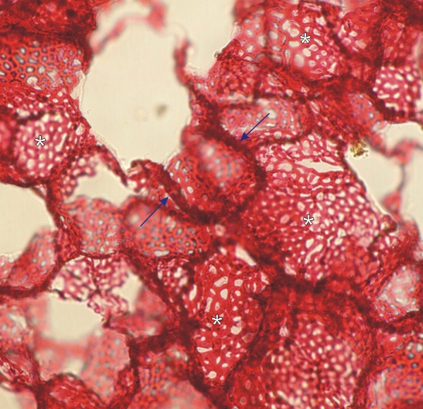
Image of a thick section of an inflated lung with the capillaries injected with a red solution.

The limits of several lung alveoli are discernible (arrows).

In the alveolar wall of several alveoli, the capillary networks are seen in face view (*).

Such a slide emphasizes the abundance of capillaries in the alveolar walls.

This preparation was made by J.S. Harrison (England), who was awarded a prize for it in 1867 in Paris!
This slide was given to Y. Clermont in 1988 by S. Emil (McGill Medical Class of 1991)
.


Magnification: ×100


We'd very much appreciate receiving your comments and suggestions!We'd very much appreciate receiving your comments and suggestions!

Creative Commons Licence The text and images of this Histology Atlas, by Yves Clermont, Michael Lalli & Zsuzsanna Bencsath-Makkai, are licensed under a Creative Commons Attribution-Noncommercial-No Derivative Works 2.5 Canada Licence and cannot be modified without the written permission of the authors. Use of any text or images must carry an acknowledgement which includes a link to the original work.


HAweb/Zs B-M -> HAbase-generated on 2013-09-13