|
||
| 10. Respiratory System | ||
| 1 2 3 4 5 6 7 8 9 10 11 12 13 14 15 16 17 18 19 20 21 22 23 24 25 | ||
| 26 27 |
| |||
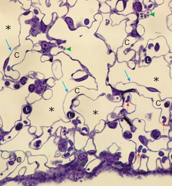 |
Semi-thin section of the lung of a rodent.
The alveolar lumina (*) and the capillary lumina (C) are indicated. The capillary lumina contain red blood cells (short red arrows), blood platelets (green arrowheads) and small lymphocytes (L). It is difficult to distinguish between alveolar type I cells and capillary endothelial cells in the alveolar wall. The purpose of this image is to emphasize the extreme thinness of the partitions (blue arrows) separating the capillary lumen from the alveolar air spaces. These partitions include the thin and indistinct cytoplasmic layers of alveolar type I cells and of endothelial cells, with a thin basement membrane in between. Such a thin wall permits the exchange of oxygen and carbon dioxide between air and the red blood cells in the blood capillaries. Stain: Toluidine blue
|
||