Contents Yves Clermont, Michael Lalli, Zsuzsanna Bencsath-Makkai
LIGHT-MICROSCOPIC HISTOLOGY ATLAS
     10. Respiratory System
  1     2     3     4     5     6     7     8     9     10     11     12     13     14     15     16     17     18     19     20     21     22     23     24     25  
  26     27  

    10.18   Lung Version française
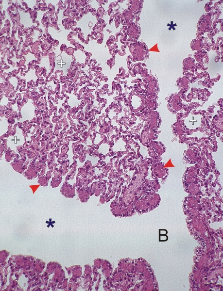
Section of the lung of a dog.

This field shows a respiratory bronchiole (B) continuous with two alveolar ducts (*).

Along the wall of the alveolar ducts, cut longitudinally, is a series of knoblike transverse sections of fascicles of smooth muscles (arrowheads). Here the knobs are close to each other, but this is due to the collapse of the lung tissue at the time of fixation.

The alveoli (+) surrounding the alveolar ducts have also collapsed for the same reason.

Stain: H–­­E
Magnification: ×300


We'd very much appreciate receiving your comments and suggestions!We'd very much appreciate receiving your comments and suggestions!

Creative Commons Licence The text and images of this Histology Atlas, by Yves Clermont, Michael Lalli & Zsuzsanna Bencsath-Makkai, are licensed under a Creative Commons Attribution-Noncommercial-No Derivative Works 2.5 Canada Licence and cannot be modified without the written permission of the authors. Use of any text or images must carry an acknowledgement which includes a link to the original work.


HAweb/Zs B-M -> HAbase-generated on 2013-09-13