|
||
| 10. Respiratory System | ||
| 1 2 3 4 5 6 7 8 9 10 11 12 13 14 15 16 17 18 19 20 21 22 23 24 25 | ||
| 26 27 |
| |||
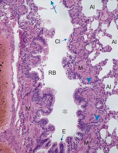 |
Section of a terminal bronchiole in the lung of a dog.
This field shows a longitudinal section of a terminal bronchiole (*) continuous with a respiratory bronchiole (RB) heading toward an alveolar duct (blue arrow at the top). The terminal bronchiole shows folds of the mucosa lined with a ciliated epithelium (E). (The cilia are not evident at this magnification.) This epithelium is next to a layer of smooth muscle fibres (M). The respiratory bronchiole is lined with a simple epithelium of pale non-ciliated cells or Clara cells (Cl) sitting on a layer of smooth muscle fibres. The wall of the respiratory bronchiole is interrupted by passageways (arrowheads) opening into the lung alveoli (Al). (Compare this image with the drawing in Figure 10.14.) Stain: HE
|
||