|
||
| 10. Respiratory System | ||
| 1 2 3 4 5 6 7 8 9 10 11 12 13 14 15 16 17 18 19 20 21 22 23 24 25 | ||
| 26 27 |
| |||
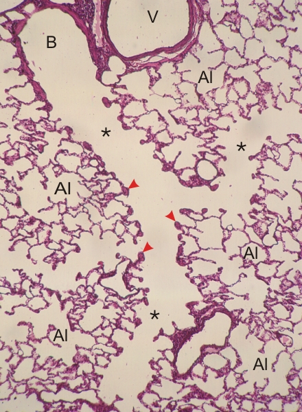 |
Section of a lung of a dog.
(Top left) This field shows a respiratory bronchiole (B) connected to branching alveolar ducts (*) of decreasing size. These ducts are delimited by knobs of smooth muscle fibres (arrowheads). In three dimensions these knobs are parts of networks as illustrated in Figure 10.14. These alveolar ducts open into groups of interconnected alveoli (Al). A large vessel (V) next to the bronchiole is also labelled. Stain: Iron hematoxylin-E
|
||