Contents Yves Clermont, Michael Lalli, Zsuzsanna Bencsath-Makkai
LIGHT-MICROSCOPIC HISTOLOGY ATLAS
     10. Respiratory System
  1     2     3     4     5     6     7     8     9     10     11     12     13     14     15     16     17     18     19     20     21     22     23     24     25  
  26     27  

    10.5   Tracheal epithelium Version française
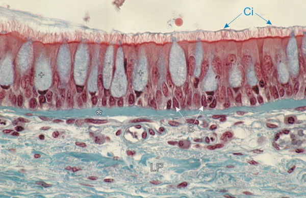
This field shows the pseudostratified columnar epithelium of the trachea.

The present epithelium is composed of ciliated columnar cells (Ci) and mucous goblet cells (+). The mucous goblet cells show a dark triangular nucleus at their base and a large pocket containing mucigen granules stained green. Many small basal cells (B) are present along the basement membrane between the basal extremities of ciliated and goblet cells.

A thick layer of type I collagen (*), stained green, separates the epithelium from the underlying connective tissue of the lamina propria (LP).

Stain: Masson’s Trichrome
Magnification: ×500


We'd very much appreciate receiving your comments and suggestions!We'd very much appreciate receiving your comments and suggestions!

Creative Commons Licence The text and images of this Histology Atlas, by Yves Clermont, Michael Lalli & Zsuzsanna Bencsath-Makkai, are licensed under a Creative Commons Attribution-Noncommercial-No Derivative Works 2.5 Canada Licence and cannot be modified without the written permission of the authors. Use of any text or images must carry an acknowledgement which includes a link to the original work.


HAweb/Zs B-M -> HAbase-generated on 2013-09-13