|
||
| 10. Appareil Respiratoire | ||
| 1 2 3 4 5 6 7 8 9 10 11 12 13 14 15 16 17 18 19 20 21 22 23 24 25 | ||
| 26 27 |
| |||
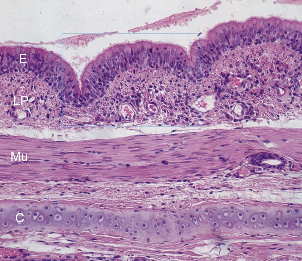 |
Coupe de la paroi dune bronche dun chien. Ce champ montre les structures qui constituent les éléments habituels de la paroi des bronches (du haut en bas):
Coloration: HÉ
|
||