Contents Yves Clermont, Michael Lalli, Zsuzsanna Bencsath-Makkai
LIGHT-MICROSCOPIC HISTOLOGY ATLAS
     10. Respiratory System
  1     2     3     4     5     6     7     8     9     10     11     12     13     14     15     16     17     18     19     20     21     22     23     24     25  
  26     27  

    10.1   Olfactory mucosa Version française
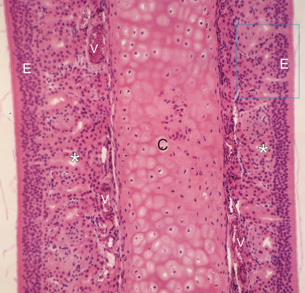
Section of the roof of the nasal cavity of a monkey.

This field shows a section of a nasal septum, and along both surfaces, the olfactory mucosa. A plate of hyaline cartilage (C) forms the core of the septum.

A pseudostratified columnar epithelium (E) lines the mucosa. In the underlying lamina propria numerous serous Bowman’s glands (*) open at the surface of the epithelium via narrow ducts.

Numerous small vessels (V) are seen next to the cartilage plate.

The framed area is magnified in Figure 10.2.

Stain: H–­­E
Magnification: ×160


We'd very much appreciate receiving your comments and suggestions!We'd very much appreciate receiving your comments and suggestions!

Creative Commons Licence The text and images of this Histology Atlas, by Yves Clermont, Michael Lalli & Zsuzsanna Bencsath-Makkai, are licensed under a Creative Commons Attribution-Noncommercial-No Derivative Works 2.5 Canada Licence and cannot be modified without the written permission of the authors. Use of any text or images must carry an acknowledgement which includes a link to the original work.


HAweb/Zs B-M -> HAbase-generated on 2013-09-13