Contents Yves Clermont, Michael Lalli, Zsuzsanna Bencsath-Makkai
LIGHT-MICROSCOPIC HISTOLOGY ATLAS
     10. Respiratory System
  1     2     3     4     5     6     7     8     9     10     11     12     13     14     15     16     17     18     19     20     21     22     23     24     25  
  26     27  

    10.6   Trachea Version française
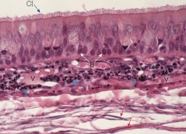
Section of a dog trachea stained to show elastic fibres.

This section shows ciliated cells (Ci), a few goblet mucous cells (+) with lightly stained mucigen granules and basal cells (B) applied to the lamina propria.

In the underlying dense lamina propria numerous cross sections of elastic fibres are stained black (vertical arrows).

In the looser connective tissue (bottom) a few longitudinal views of elastic fibres (red arrows) are seen in addition to the acidophilic collagen fibres.

Small vessels (V) are also labelled.

Stain: Iron hematoxylin-E
Magnification: ×500


We'd very much appreciate receiving your comments and suggestions!We'd very much appreciate receiving your comments and suggestions!

Creative Commons Licence The text and images of this Histology Atlas, by Yves Clermont, Michael Lalli & Zsuzsanna Bencsath-Makkai, are licensed under a Creative Commons Attribution-Noncommercial-No Derivative Works 2.5 Canada Licence and cannot be modified without the written permission of the authors. Use of any text or images must carry an acknowledgement which includes a link to the original work.


HAweb/Zs B-M -> HAbase-generated on 2013-09-13