Contents Yves Clermont, Michael Lalli, Zsuzsanna Bencsath-Makkai
LIGHT-MICROSCOPIC HISTOLOGY ATLAS
     10. Respiratory System
  1     2     3     4     5     6     7     8     9     10     11     12     13     14     15     16     17     18     19     20     21     22     23     24     25  
  26     27  

    10.26   Lung Version française
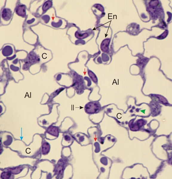
Semi-thin section of the lung of a rodent.

The following structures are labelled:

  • Alveolar lumina (Al)
  • Capillary lumina (C)
  • Red blood cells (red arrows)
  • Blood platelets (green arrowhead)
  • Nucleus of one alveolar type II cell (II)
  • Nuclei of two endothelial cells (En)

As in Figure 10.25, this image emphasizes the thinness of the wall (blue arrow) separating the air space of the alveolar lumen and the blood space of the capillaries.

Stain: Toluidine blue
Magnification: ×1100


We'd very much appreciate receiving your comments and suggestions!We'd very much appreciate receiving your comments and suggestions!

Creative Commons Licence The text and images of this Histology Atlas, by Yves Clermont, Michael Lalli & Zsuzsanna Bencsath-Makkai, are licensed under a Creative Commons Attribution-Noncommercial-No Derivative Works 2.5 Canada Licence and cannot be modified without the written permission of the authors. Use of any text or images must carry an acknowledgement which includes a link to the original work.


HAweb/Zs B-M -> HAbase-generated on 2013-09-13