Contents Yves Clermont, Michael Lalli, Zsuzsanna Bencsath-Makkai
LIGHT-MICROSCOPIC HISTOLOGY ATLAS
     10. Respiratory System
  1     2     3     4     5     6     7     8     9     10     11     12     13     14     15     16     17     18     19     20     21     22     23     24     25  
  26     27  

    10.3   Trachea Version française
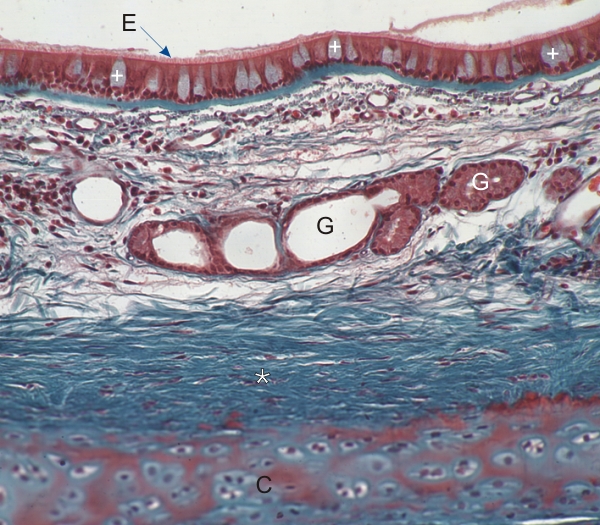
Section of the trachea of a monkey.

The lumen of the trachea (above) is lined with a pseudostratified columnar epithelium (E) composed of intensely stained ciliated cells and lightly stained goblet mucous cells (+).

Underlying this epithelium, the lamina propria composed of loose connective tissue, contains the profile of a sero-mucous gland (G).

Deeper in the wall of the trachea, a layer of dense connective tissue stained green (*) is next to the plate of hyaline cartilage (C).

Stain: Masson’s Trichrome
Magnification: ×150


We'd very much appreciate receiving your comments and suggestions!We'd very much appreciate receiving your comments and suggestions!

Creative Commons Licence The text and images of this Histology Atlas, by Yves Clermont, Michael Lalli & Zsuzsanna Bencsath-Makkai, are licensed under a Creative Commons Attribution-Noncommercial-No Derivative Works 2.5 Canada Licence and cannot be modified without the written permission of the authors. Use of any text or images must carry an acknowledgement which includes a link to the original work.


HAweb/Zs B-M -> HAbase-generated on 2013-09-13