|
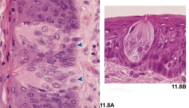
Sections of the crypts of the circumvallate papillae from the tongue.
Shown here are longitudinal sections of taste buds (*). Taste buds are composed of several types of cells. Some of them are fusiform and their apical processes reach a small narrow space or pore opening at the surface of the stratified epithelium. Some taste pores are indicated by arrows in the two images. In addition to the fusiform cells of the buds in Figure 11.8A, some small vacuolated basal cells (arrowheads) are visible close to the underlying connective tissue.
Some fine nervous terminals (not visible here) enter these buds and establish close contacts, or synapses, with some fusiform cells.
Stain: HE
Magnification: ×900
|